Breadcrumb
Stable mandibular landmarks in adolescent patients
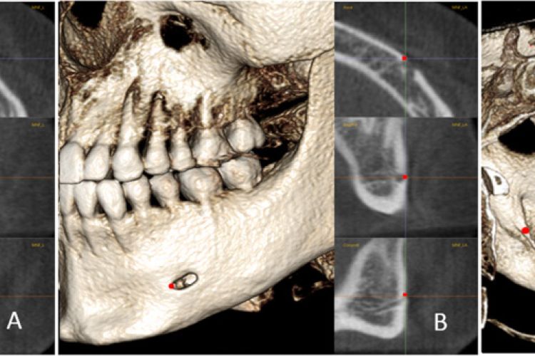
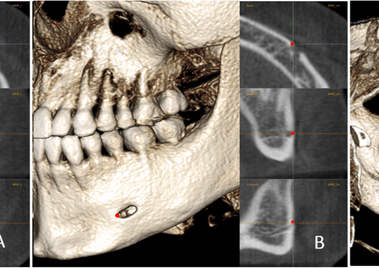
The positions of MF, MFA, and MdF on the left side: a, MF is located in center of the mental foramen; b, MFA is the most anterior point of the mental foramen; c, MdF is located in the center of the mandibular foramen. It is placed on the first slice where the canal shows a complete circle from superior to inferior
The three-dimensional stable mandibular landmarks in patients between the ages of 12.5 and 17.1 years
What is it?
A study of 20 growing patients between the ages of 12.5 and 17.1 years.
What problem does it aim to solve?
The goal is to identify the most stable mandibular landmarks in growing patients using three-dimensional images.
How does it work?
The sample was comprised of two cone-beam computed tomography (CBCT) scans taken about 4.6 years apart in 20 adolescents. After head orientation, landmarks were located on the chin, internal symphysis of the mandible, and mandibular canals. The linear distance change between the landmarks was measured on each CBCT to test stability over time and to determine how reliable the landmarks were.
What are the real-world implications?
Better understanding of how the jaw develops in adolescence.
“The centers of the mandibular foramina and the starting points of the mandibular canal underwent significant changes in the transverse and sagittal dimensions… We also introduced new stable landmarks for mandibular superimposition. Pog, landmarks on the inferior part of the internal symphysis (C, D, and E), and the mental foramen appear relatively stable, and thus, can be used in mandibular regional superimpositions.”
What are the next steps?
Further study. In a future study, we plan to investigate 3D growth of the mandibular canals and to explore more stable structures based on the stable landmarks that we have identified to improve the voxel-based superimposition method in growing subjects. In addition, further validation studies against other samples are required for the suggested stable landmarks in the present study.
Source
Chen, G., Al Awadi, M., Chambers, D.W. et al. The three-dimensional stable mandibular landmarks in patients between the ages of 12.5 and 17.1 years. BMC Oral Health 20, 153 (2020).
Authors
Gui Chen, Department of Orthodontics, Peking University School and Hospital of Stomatology, National Engineering Laboratory for Digital and Material Technology of Stomatology, Beijing Key Laboratory of Digital Stomatology
Mona Al Awadi, Department of Orthodontics, Arthur A. Dugoni School of Dentistry
David William Chambers, Department of Orthodontics, Arthur A. Dugoni School of Dentistry
Manuel O. Lagravère-Vich, Associate Professor of Department of Dentistry, Faculty of Medicine and Dentistry, University of Alberta, Edmonton
Tianmin Xu, Department of Orthodontics, Peking University School and Hospital of Stomatology, National Engineering Laboratory for Digital and Material Technology of Stomatology, Beijing Key Laboratory of Digital Stomatology
Heesoo Oh, Department of Orthodontics, Arthur A. Dugoni School of Dentistry